Electric catfish with unusual swelling
- Thread starter DemiGorgon
- Start date
You are using an out of date browser. It may not display this or other websites correctly.
You should upgrade or use an alternative browser.
You should upgrade or use an alternative browser.
I wonder that, too. I'm following odball's treatment protocol for bloat (the epsom salt variation), and she's hanging in there, but not really improving. So far. The swelling has spread some, to include some area cranial of the anal pore, but still not looking like abdominal cavity involvement. Treating with praziquantel, as well, and planning to treat for parasitic nematodes (wont hurt, could be at least part of the problem); on my way to lfs now. It'll be interesting, since I've never given oral liquids to a fish before, and she still won't eat. Wish me luck! And if anyone has any new thoughts, I'd love to hear them! Or advice on how to administer oral liquids to an e-cat.
It doesn't look like a digestive issue.
I personally would not medicate the fish because IDK what I'd be doing. It'd be a gamble - you may luck out and the med(s) may save the fish in one way or another (the chances of this are slim to none) or you may pile additional stress on the fish that even if the medicine was right or wrong or unneeded, it won't matter as the fish may not handle so much stress (these chances are significant).
There is a way to force-administer an oral medicine to a fish but a liquid kind would be pretty hard plus this is an electric fish - it will zap anyone attempting this repeatedly until it's let go. It's a pinnacle of stressful handling of any fish.
You can gather an idea how this is done through this thread and videos and those linked in there (there are some others too) http://www.planetcatfish.com/forum/viewtopic.php?f=7&t=37031&hilit=english
I agree that it doesn't look digestive, so I didn't do the wormer. I continued the Abx and water changes, but the edema continued to spread forward. Water quality was checked at least every other day, with a liquid test kit, and was always good.
Sparky died this morning.
In the interest of increasing our collective knowledge, I did a necropsy (after the first round of crying, obviously).
I found a few obviously unusual things, and one that I'm not sure if it's normal or abnormal.


Sparky died this morning.
In the interest of increasing our collective knowledge, I did a necropsy (after the first round of crying, obviously).
I found a few obviously unusual things, and one that I'm not sure if it's normal or abnormal.


Aaaahhhh! I'm so sorry; I clicked "upload as thumbnails", but it didn't!I agree that it doesn't look digestive, so I didn't do the wormer. I continued the Abx and water changes, but the edema continued to spread forward. Water quality was checked at least every other day, with a liquid test kit, and was always good.
Sparky died this morning.
In the interest of increasing our collective knowledge, I did a necropsy (after the first round of crying, obviously).
I found a few obviously unusual things, and one that I'm not sure if it's normal or abnormal. View attachment 1213665 View attachment 1213666 View attachment 1213667 View attachment 1213668
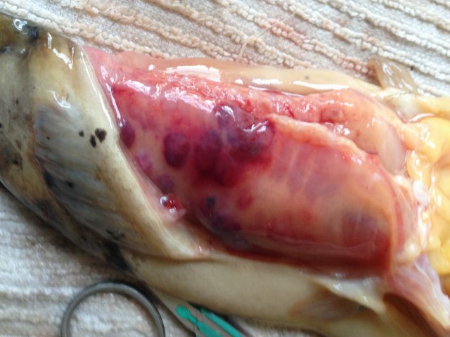
Pic 1 is the bubbly, bruise-looking spots she had on her tail muscle. It was superficial, only; the muscle beneath was normal-looking. When I cut into the spots, I was able, with some effort, to express a little light-colored tissue with the consistency of fat tissue. I prepped a slide of it, so I can look at it under the microscope tomorrow morning.
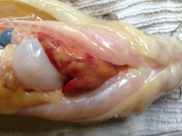
Pic 2 is gall- and swimbladders on the left, yeah?, but the liver and its blotchy, yellowish appearance is what I was documenting.
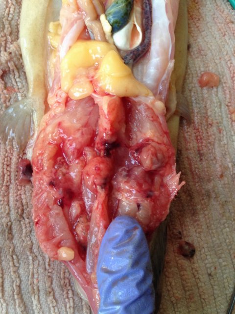
Pic 3 is a view of her spine, through the swimming muscle that is caudal to the anal pore, so we are looking from the ventral side. Not sure what this hollowed-out area in the middle of her muscle is. Ruptured tumor? I would guess abscess, except there was no fluid: pus, blood or serous, inside that hollow. But the disorganized-looking tissue looks like an abscess does when it's been cleaned out. Or a tumor does when it outgrows itself.
That was the epicenter, though, of the swelling.
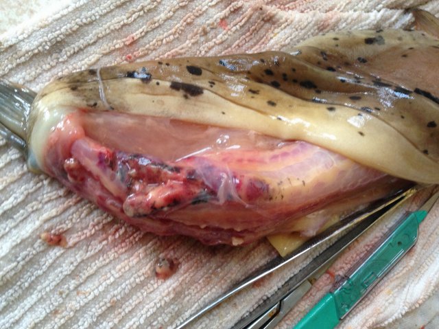
Pic 4 is the part that I'm not sure if it's normal or not; I wish I had snapped a pic before I cut into it. She seemed to have a channel or tube running the length of her dorsal spine. It was full of a gelatinous tissue that I've not encountered before. Over the muscular caudal end of her body, it looked bruised and had black mottling, although you could make a case that it matches the blotching of her skin, and I'm assuming the bruising was due to pressure from the edema.
The swelling was whole-body by the end, but there was no pocket of fluid of any kind, nor was her body cavity filled with fluid; it was diffused throughout all her tissue.
Her gut was empty (you can see a little in pic 2). I ran it as best as an amateur can, and didn't see anything unusual -- like parasites or obstruction.
Anything look like something anyone knows more about? I'd love to hear your thoughts! If not, well, this is what I found, and maybe it will be useful to someone one day.


