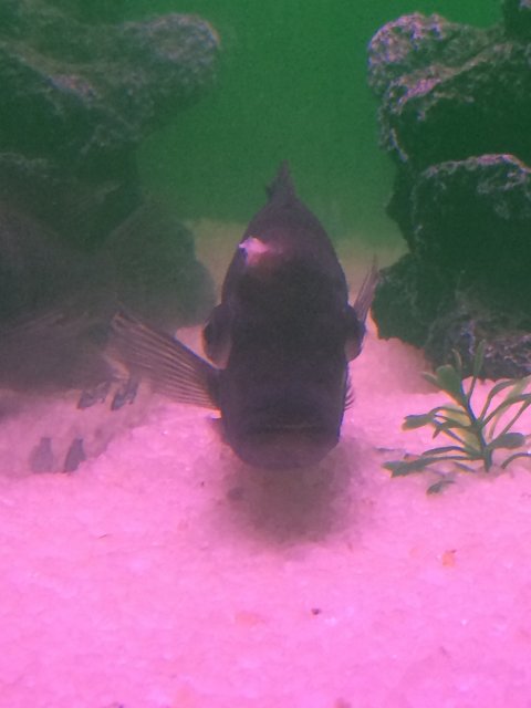
My frontosa is having cotton wool fungal infection pls help

Sumit Panigrahy

Feeder Fish
MFK Member
Jun 2, 2016
29
3
3
26
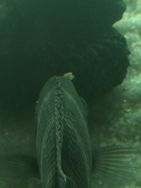
He is suffering what i think is cotton wool disease. Yesterday when i noticed the cotton growth on his bump i separated him and applied anti fungal medication the wool sheded and again reappeared today . He is not eating anything and is going weak this way he will starve before i treat him . Pls help what can i do to make him healthy.image.jpeg image.jpeg
 

Wailua Boy

Potamotrygon
MFK Member
Jan 2, 2015
2,752
1,315
164
Hawaii
I didnt notice the bulging eyes at first, now im starting to think water quality is an issue
 

Wailua Boy

Potamotrygon
MFK Member
Jan 2, 2015
2,752
1,315
164
Hawaii
The spot on his head doesnt look bacterial in nature but the bulging eyes may indicate a severe bacterial conditon
 

Charney

The Fish Doctor
Staff member
Moderator
MFK Member
Nov 15, 2005
3,597
665
150
40
Somerville NJ
could you get a better picture of the wound. Could be bacterial either primary or secondary
 

Wailua Boy

Potamotrygon
MFK Member
Jan 2, 2015
2,752
1,315
164
Hawaii
On second thought, I'm leaning towards a lympho infection as bulging eyes can be a symptom; heres some background on the disease:


  • Home
  • FAQs & Help
  • Local Offices
  • IFAS Bookstore
  • Advanced Search
  • Download PDF
    Publication #FA181
    2
    Lymphocystis Disease in Fish1
    Roy P. E. Yanong, VMD2

    Figure 1.
    African cichlid with lymphocystis nodules on fins.


    Credit:
    Roy Yanong


    [Click thumbnail to enlarge.]


    Figure 2.
    Clownfish with lymphocystis nodules on fins and body.


    Credit:
    Roy Yanong


    [Click thumbnail to enlarge.]


    Pop-eye (exophthalmia; marked protrusion of the eye) has also been seen and results from lymphocystis masses in or behind the eye pushing the eyeball out.

    Fish with lymphocystis normally do not behave differently from uninfected fish in the same group. However, if there are large numbers of nodules on the body or fins, or if they are covering a large portion of the gills, these may alter swimming or breathing patterns.

    Figure 3.
    Wet mount. Microscopic view of nodules on fins of blue gourami.


    Credit:
    Roy Yanong


    [Click thumbnail to enlarge.]


    Figure 4.
    Wet mount. Close up of enlarged lymphocystis infected cell on fins of a clownfish.


    Credit:
    Roy Yanong


    How do I tell if my fish has lymphocystis?
    In addition to seeing the wart-like growths on the fins, skin, or gills, a wet mount (fresh) biopsy of these growths (careful removal of one of these growths from the skin, fins, or gills) should be examined under the microscope. A strong tentative diagnosis of lymphocystis can be made based on seeing round to oval, grape, or balloon like structures, often in clumps (see figures 3 and 4).

    Fish may also be infected with lymphocystis without having obvious lesions that are visible with the naked eye. These fish may have smaller infected cells or may have one or more internal organs infected (e.g., inside the eye, spleen, or other organs). Lymphocystis, in these cases, may only be seen after microscopic examination of a wet mount (fresh) biopsy of suspected infected tissues, or by using more advanced techniques such as histopathology, electron microscopy, or molecular methods.

    For an accurate diagnosis, work with a fish health professional or fish disease diagnostic laboratory if you suspect your fish have lymphocystis.

    Can lymphocystis be confused with any other diseases?
    Yes. In early stages of the disease, the nodules may be relatively small and may be confused with the parasites Ichthyophthirius multifiliis(Ich, freshwater white spot disease), Cryptocaryon irritans (marine white spot disease), digenean trematodes ("grubs"), or heavy infection with Epistylis (a stalked, ciliated, protistan parasite). Lymphocystis nodules may also be confused with epitheliocystis (an intracellular bacterial disease), clumps of fungus, or mucus tags. Some skin neoplasias (cancers) may also be confused with lymphocystis.

    Because of the potential confusion with other disease agents, diagnosis of lymphocystis should be accomplished by a qualified fish health professional to rule out these other potential causes.

    What factors contribute to development and spread of lymphocystis disease?
    Lymphocystis is spread by fish-to-fish contact or contact with infected tissues. External trauma from spawning, aggression, parasites, or handling can facilitate infection and spread. In addition, crowding, shipping, and other stressors appear to trigger disease outbreaks. Lymphocystis does not appear to be spread by vertical transmission (i.e., from parents to offspring via infection of eggs or sperm).

    Although all contributing factors are not known, there do appear to be different genetic variants or possibly "species" of Lymphocystivirus. Each of these affects a different group of fish. Because the virus that causes lymphocystis has been difficult to work with, experts have only officially assigned one Lymphocystivirus species, Lymphocystis disease virus 1 (LCDV-1). Several other variants have been suggested and may be determined to be separate species (including LCDV-2, LCDV-C), but these have not yet been officially accepted by all virus experts.

    Based on clinical observations of mixed species groups and other anecdotal evidence, there may be additional strain variations and/or differences in individual fish species sensitivity to infection. In mixed natural or aquarium populations of fish, typically only one or two species will be infected. However, in experimental settings, multiple species can be infected by direct exposure to infected tissues.

    What happens to infected fish?
    Lymphocystis is usually a self-limiting disease, meaning that, in most cases, the lesions will clear up after a few weeks in warmwater fish species (up to 6 weeks in cool or coldwater species). However, because of the unsightly growths, fish normally cannot be sold during this time. If a fish has numerous lesions and/or lymphocystis nodules cover a large portion of important organs (e.g., the gills), there may be some impairment and greater chance of secondary infections by bacteria, parasites, or fungi that can now more easily infect the fish and help contribute to mortalities.

    Can fish with lymphocystis be treated?
    Currently, there is no good treatment that will speed up recovery from this disease. Most often, the disease must run its course in an affected fish. Fortunately most cases of lymphocystis in warmwater fish will resolve on their own after a few weeks, as long as husbandry is good (good water quality/chemistry, good nutrition, correct population densities, optimal social groups) and as long as other stressors have been eliminated. Although it is not an ideal solution, at present, the best option is to hold fish for several weeks (longer for cool and cold water fish) until the lesions have cleared.

    Because lymphocystis disease tends to develop in parts of tissues that are less exposed to the immune system (e.g., at the periphery, away from blood vessels that carry immune cells) and in tissues with a thick hyaline membrane that may "hide" these cells, an immune response normally does not develop until after cells have burst and released virus. There is some evidence that fish infected with lymphocystis will develop less pronounced lesions if they are re-infected or if lesions recur.

    Culling (removing) infected fish from a population may help reduce overall loads of virus in the system as well as infection rate, but it is difficult to cull all affected fish because some infections may not be visible with the naked eye, and many less severely infected fish may remain in the population to potentially infect other fish. Culling, therefore, may not be as effective a measure for aquacultured populations as for hobbyists or display aquaria. Culled fish can be isolated until their lesions resolve, but lesions may recur. If they do, they will most likely be less severe.
 

Charney

The Fish Doctor
Staff member
Moderator
MFK Member
Nov 15, 2005
3,597
665
150
40
Somerville NJ
a better picture would help. With lympho you typically don't have an ulcerated lesion under it which If feel I can kind of see in the picture. Hard to say for sure
 
zoomed.com
hikariusa.com
aqaimports.com
Store